Laboratory of Biological Inorganic Chemistry, Department of Chemistry, Graduate School of Science, Osaka Metropolitan University
& 自然科学研究機構 分子科学研究所 特別研究部門(生命・錯体分子科学研究領域領域)Institute for Molecular Science, National Institute of Natural Sciences
研究内容 Research projects
生命の無機化学:生体内金属動態の理解と制御/細胞内微量金属の新しい定量法の開発Biological Inorganic Chemistry for Life: Understanding and Controlling Metal Dynamics in Living Systems / Novel Quantitative Analysis of Trace Metals in Cells
からだの中で「金属」は重要なはたらきをしています!
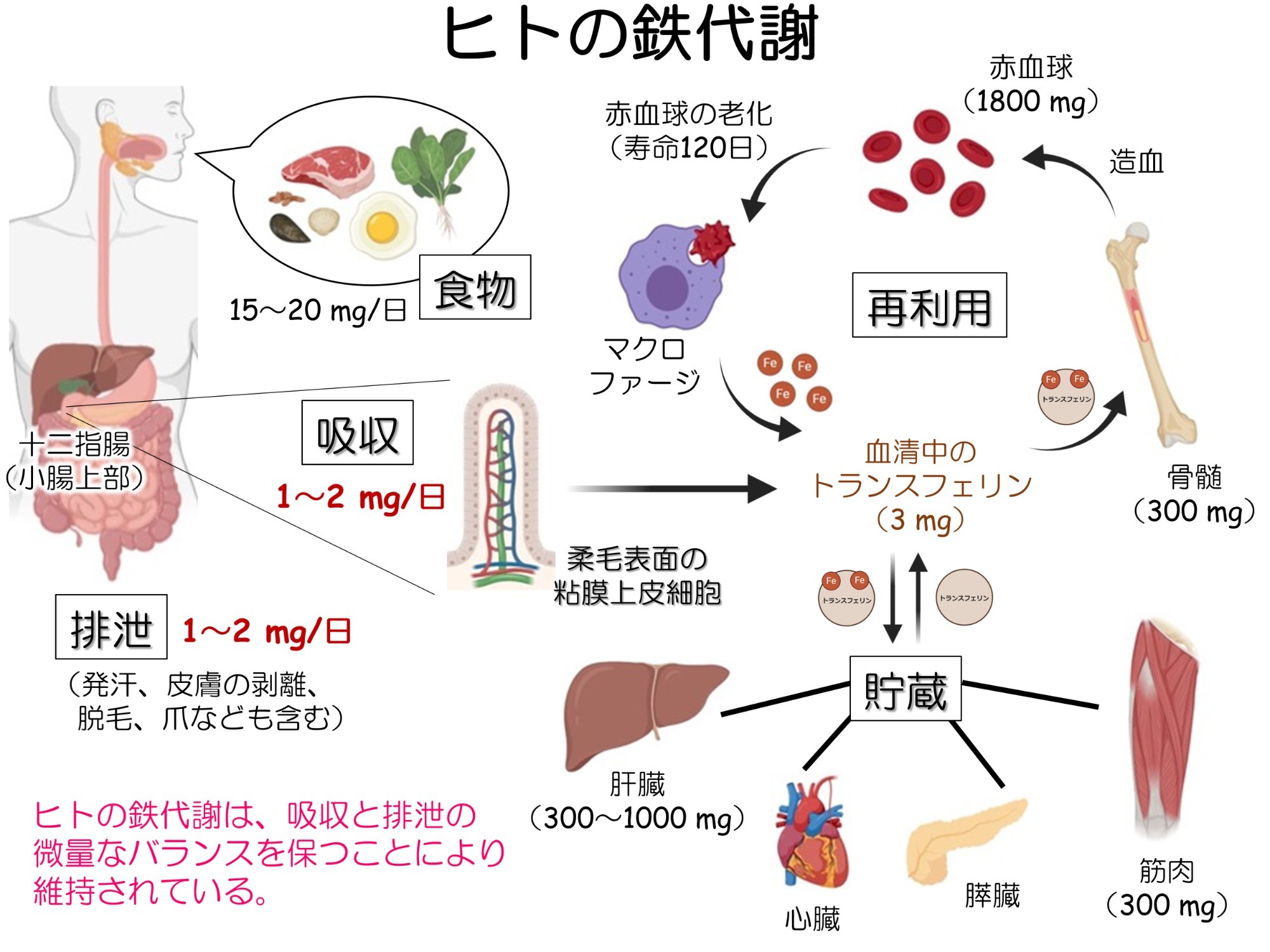
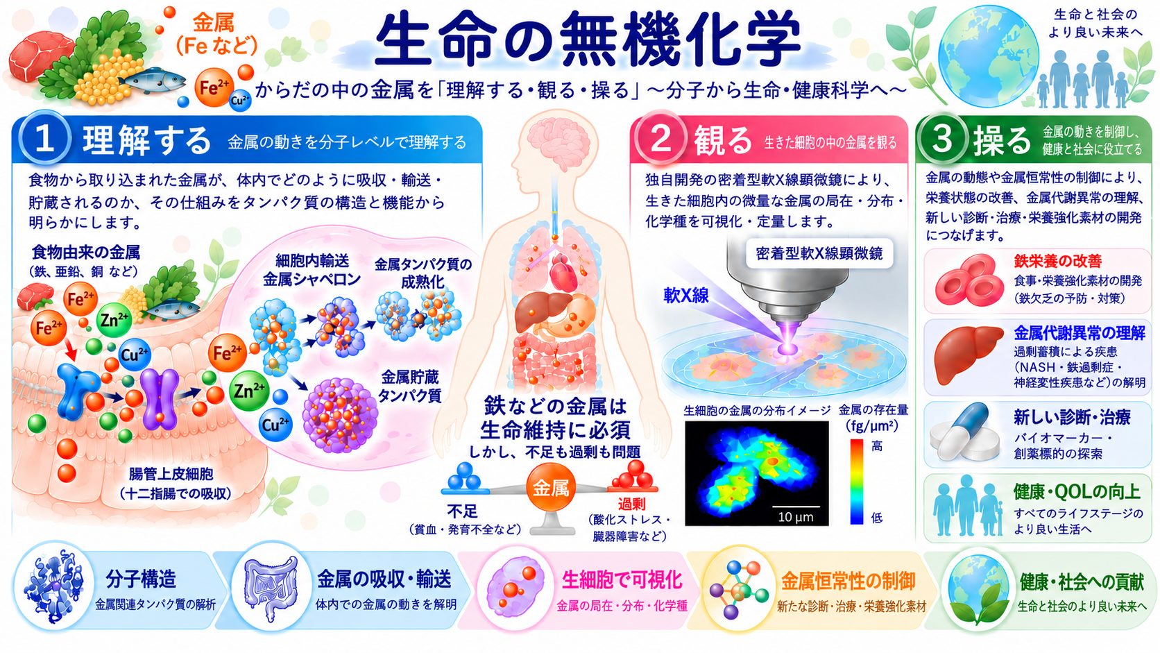
ヒトの健康維持に鉄分の摂取が有効であることは、古くから経験的に知られてきました。しかし、生体内の鉄の動態(吸収・感知・輸送・貯蔵など)の機序や他の金属との識別選択メカニズムについては、未だに分子科学的な理解が進んでいません。そのため、鉄をはじめとする生体金属の代謝異常による疾病(たとえば、非アルコール性脂肪肝炎・ALSやアルツハイマー病などの神経変性疾患)に対する特効薬はありません。また市販のサプリメントや鉄剤があるにもかかわらず、世界人口の約30%に鉄欠乏の症状がみられ、生活の質(QOL)を低下させる要因にもなっています。私たちは、生体内の鉄をより深く理解し、その知見を将来的にはヒトの健康やQOLの向上に活かしたいと考え、鉄栄養素の選択的な吸収・濃度感知・細胞内輸送などを担う様々なタンパク質の立体構造を最先端の手法を用いて解明するとともに、細胞の機能との関連性について探究しています。さらに、生きている細胞の中に存在する各種金属の分布を化学種ごとに可視化できる「生細胞丸ごと金属マッピング法(軟X線顕微鏡法)」の開発にも取り組んでいます。からだの中ではたらく「金属」は多すぎても少なすぎても病気になります。栄養素として金属を利用するための仕組みを詳しく調べることは、病気を治すための「新しい医薬品のデザイン」、食品から安全に効率よく金属栄養素を摂取するための「栄養強化素材の開発」、さらには金属代謝異常を起因とする疾病の「新しい診断技術の開発」につながります。
Metals play important roles in the body!
Some metals function as essential nutrients, playing critical roles in regulating vital physiological functions but in excess they can produce toxic effects. Those contained in food and drink are reduced in toxicity by binding to a wide variety of proteins. These metals are used as active centers of enzymes that carry out functions essential to sustaining life, e.g. transport and storage of oxygen, energy production, gene synthesis. However, the molecular mechanisms for the series of proteins controlling metal dynamics in the body (absorption, sensing, transport, storage, and excretion) and their selectivity for individual metals, remain to be elucidated. Our research focuses on "iron", which is the most important metal among the essential metals for sustaining life. Our work centers on various proteins that play a role in the selective absorption, sensing, and intracellular transport of iron in food. The goal is to elucidate the series of molecular mechanisms for iron dynamics in humans and to decipher "tactics" of organisms to utilise iron, which has both properties of nutrient and poison in physiological processes. We explore their functions in human cells.
生細胞内の金属を可視化する ~密着型軟X線顕微鏡の開発による「生細胞丸ごと金属マッピング法」の確立 ~
Visualizing Intracellular Metals in Living Cells: Establishing a “Whole-Live-Cell Metal Mapping” Method Using Contact-Type Soft X-ray Microscopy
生きている細胞において、生体分子が「どこに、どれだけ存在しているのか?」を知ることは、生命現象を理解するうえで非常に重要です。現在、細胞内を可視化する技術として、蛍光顕微鏡、ラマン顕微鏡、質量分析イメージングなど、様々な手法が利用されています。しかし、生細胞内の金属の電子状態に着目し、それらの化学状態や酸化還元状態を区別して可視化する手法は未だ確立されていません。そこで私たち研究室では、生細胞中に微量に存在する金属の局在と分布を定量的に可視化する新しい技術として
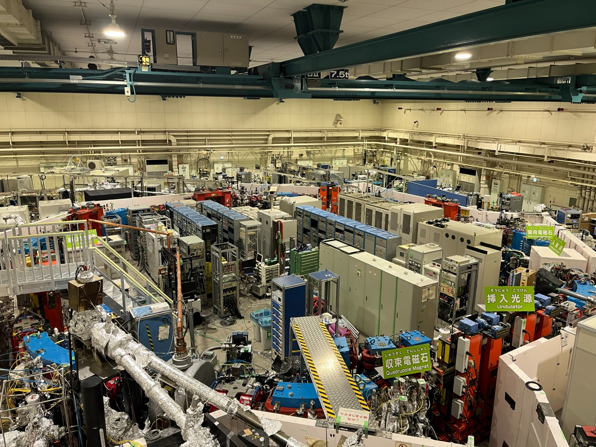
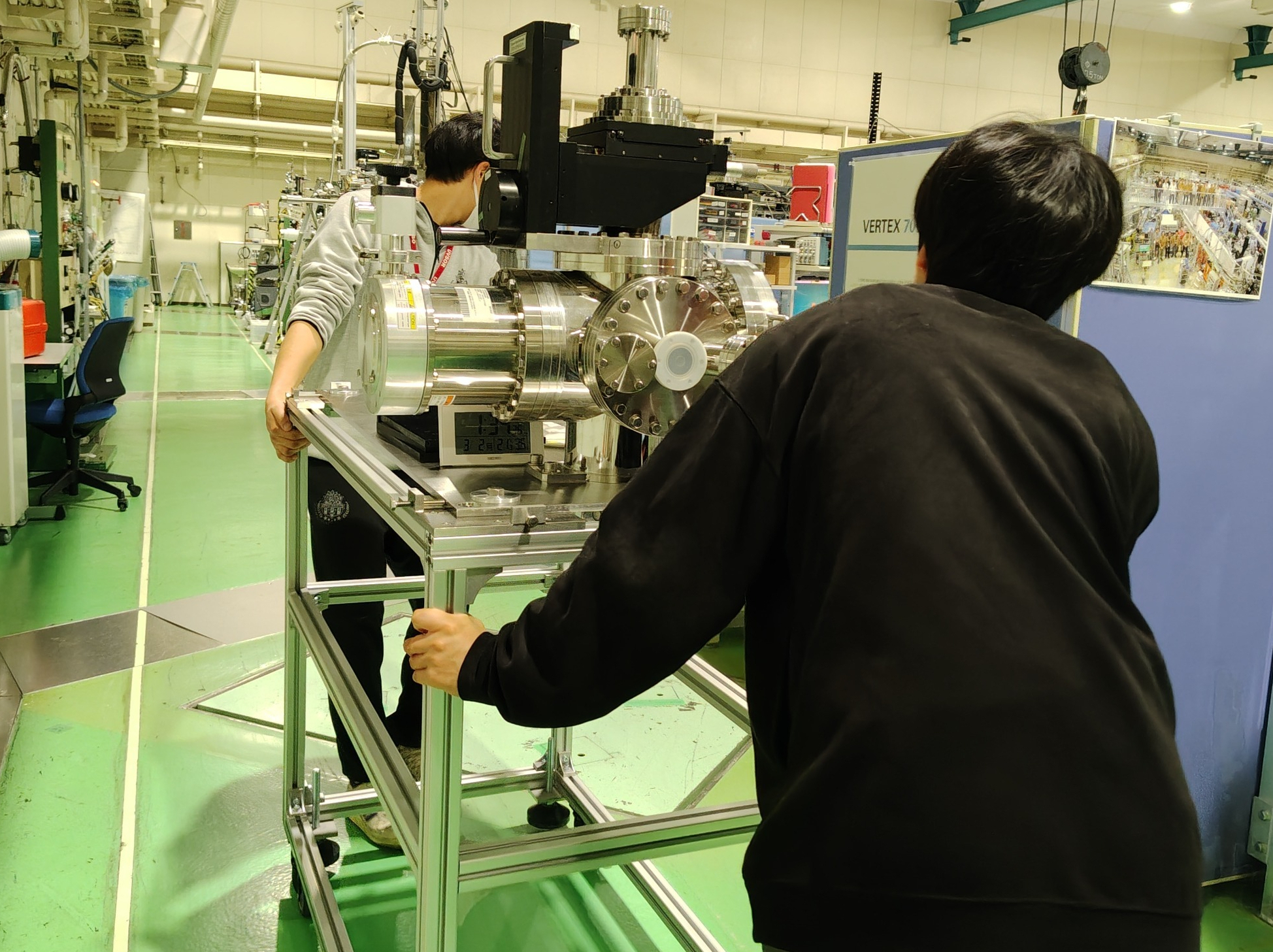
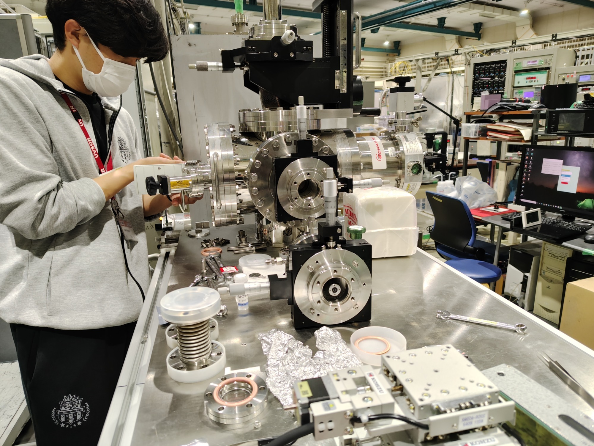
「密着型軟X線顕微鏡」の開発を自然科学研究機構 分子科学研究所にある極端紫外光研究施設UVSORにて進めています。
軟X線は、一般的なX線結晶構造解析などで用いられる硬X線よりも低いエネルギー領域のX線です。この領域では、炭素、窒素、酸素などの生体を構成する主要な元素に加えて、鉄をはじめとする様々な生体微量金属に固有の特徴的なX線吸収がみられます。このような元素選択性を利用することで、特定の元素を選択して可視化することができます。さらに、吸収エネルギーのわずかな違いを詳しく解析することで、将来的には元素の存在量だけでなく、酸化状態や化学状態の違いを識別して可視化することを目指しています。
私たちが開発した「密着型軟X線顕微鏡」の大きな特徴は、複雑な装置システムを必要とせず、生の試料をそのまま試料セルに貼り付けるだけで、試料中に存在する微量元素を直接イメージングできる点です。現在、鉄を中心とした生体微量金属の定量イメージングに加え、より広い視野で細胞や組織を観察し、複数の微量金属を化学状態ごとに可視化する技術への展開を目指しています。
生きている細胞内に存在する多様な微量元素について「どの元素が、どこに、どれだけ、どのような状態で存在するのか?」を観るだけで知ることができる「生細胞丸ごとマッピング法」の確立を目指しています。装置の小型化や解析技術の高度化を進めることで、生命科学・材料科学の研究だけでなく、将来的には疾患の診断や病態解析など、医療分野への応用につながる新しいイメージング技術へと発展させたいです。
Understanding where biomolecules are located and how much of them is present in living cells is essential for elucidating biological processes. Various imaging techniques, including fluorescence microscopy, Raman microscopy, and mass spectrometry imaging, are currently used to visualize the interior of cells. However, methods that can visualize metals in living cells while distinguishing their chemical and redox states based on their electronic structures have yet to be established.
To address this challenge, our laboratory is developing a new technique called contact-type soft X-ray microscopy at the UVSOR Synchrotron Facility. This technique enables the quantitative visualization of the localization and distribution of trace amounts of iron in living cells.
Soft X-rays have lower energies than the hard X-rays commonly used in techniques such as X-ray crystallography. In the soft X-ray energy range, characteristic X-ray absorption features can be observed not only for major biological elements such as carbon, nitrogen, and oxygen, but also for various trace metals, including iron. By taking advantage of this elemental selectivity, specific elements can be selectively visualized. Furthermore, detailed analysis of subtle differences in X-ray absorption energies may eventually enable us to distinguish and visualize not only the amounts of individual elements but also differences in their oxidation and chemical states.
A major advantage of our contact-type soft X-ray microscope is its simple measurement principle, which does not require a complex instrumental setup. Biological samples can be placed directly in a sample cell, allowing trace elements within the sample to be imaged directly. We are currently working on quantitative imaging of biological trace metals, particularly iron, while also expanding the technique to observe cells and tissues over larger fields of view and to visualize multiple trace metals according to their chemical states.
Ultimately, our goal is to establish a “whole-cell mapping” technique that reveals, simply by imaging, “which elements are present, where they are located, how much is present, and in what chemical states” within living cells. By further miniaturizing the instrumentation and advancing the analytical methods, we aim to develop this technology into a new imaging platform with applications not only in life and materials sciences, but also, in the future, in medical fields such as disease diagnosis and pathological analysis.
生命は「鉄」をどのように制御しているのか? ~ 細胞内鉄動態の分子メカニズムの解明 ~
How Does Life Control Iron? — Molecular Mechanisms of Intracellular Iron Trafficking and Homeostasis
鉄は、生命を維持するために欠かすことのできない金属元素の一つです。食物から摂取した鉄は主に十二指腸で吸収され、様々なタンパク質に受け渡されながら、エネルギー産生をはじめとする重要な生命活動に利用されています。一方で、Fe²⁺は高い化学反応性をもつため、細胞内に過剰に存在するとフェントン反応を介して強力な活性酸素種であるヒドロキシラジカルの生成を促し、細胞に深刻なダメージを与える危険性があります。そのため、生体内では鉄の「量」だけでなく「細胞内のどこで、どのような状態で、受け渡されるのか?」を精密に制御する必要があります。
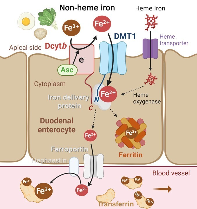
十二指腸の粘膜上皮細胞では、食物由来の鉄は腸管腔側の細胞膜に存在する鉄還元酵素DcytbによってFe³⁺からFe²⁺へと還元された後、二価金属トランスポーターDMT1を介して細胞内へ取り込まれます。取り込まれた鉄は、そのまま細胞内を自由に移動するのではなく、鉄の受け渡しを担うタンパク質を介して適切な場所へと運ばれると考えられています。鉄の一部は貯蔵タンパク質Ferritinに安全な形で蓄えられ、また一部は血管側へ輸送・放出された後、血液中のTransferrinと結合して全身を巡ります。しかし、この一連の鉄動態において、反応性の高いFe²⁺がどのように安全に受け渡され、その毒性を回避しながら細胞内の鉄恒常性が維持されているのか等の分子機構については、多くの謎が残されています。
私たちの研究室では、タンパク質間で鉄の受け渡しを仲介する「金属輸送シャペロンタンパク質」に着目しています。このタンパク質が、必要な場所へ必要な量の鉄を安全に届けることで、細胞内の鉄恒常性を維持できるようになります。タンパク質の立体構造解析や相互作用解析などを通して「鉄イオンがどのようにタンパク質からタンパク質へと受け渡されるのか?」を原子から細胞のレベルで明らかにし、生命が鉄の有用性を引き出しながら、その毒性を巧みに回避する仕組みの全容解明を目指しています。
Iron is one of the essential metal elements required to sustain life. Iron obtained from the diet is absorbed primarily in the duodenum and transferred among various proteins, through which it is utilized for vital biological processes, including energy production. At the same time, Fe²⁺ is highly reactive. When present in excess within cells, Fe²⁺ can promote the Fenton reaction, generating hydroxyl radicals, highly reactive oxygen species that can cause severe cellular damage. Therefore, living organisms must tightly regulate not only the amount of iron but also where it is located and how it is transferred within cells.
In duodenal epithelial cells, dietary iron is first reduced from Fe³⁺ to Fe²⁺ by the ferric reductase Dcytb, located on the apical membrane facing the intestinal lumen, and is then taken up into the cells via the divalent metal transporter DMT1. Once inside the cell, iron is thought to be delivered to appropriate destinations by proteins involved in iron transfer, rather than diffusing freely throughout the cytosol. Some of the iron is safely stored in the iron-storage protein ferritin, while another portion is transported toward the basolateral side and released into the bloodstream, where it binds to transferrin and is distributed throughout the body. However, many questions remain regarding how highly reactive Fe²⁺ is safely transferred through this series of processes and how intracellular iron homeostasis is maintained while avoiding iron-induced toxicity.
Our laboratory focuses on metal chaperone proteins that mediate the transfer of iron between proteins. By safely delivering the appropriate amount of iron to where it is needed, these proteins are thought to play an important role in maintaining intracellular iron homeostasis. Through structural analyses of proteins and protein complexes, together with studies of protein–protein interactions, we aim to elucidate how iron ions are transferred from one protein to another, from the atomic to the cellular level. Ultimately, our goal is to reveal the molecular mechanisms by which living systems harness the essential functions of iron while skillfully avoiding its inherent toxicity.